Background: Popliteal fossa cysts (PFCs aka Baker's cysts) are synovial cysts of the knee joint that can be symptomatic or asymptomatic and incidentally identified on ultrasound. Whether PFCs are associated with deep vein thrombosis (DVT) is unknown. Possible mechanisms for an association include direct compression of the popliteal vein, indirect compression on the popliteal vein with leg flexion, adjacent inflammation of the cyst, or relative immobility due to underlying joint disease itself.
Methods: Lower extremity venous Duplex ultrasound radiology reports from the inception of electronic archiving through 11/14/2019 were evaluated across the Mayo Clinic Enterprise (Rochester MN, Jacksonville, FL, Scottsdale AZ, and Mayo Clinic Health System) in patients >18 years of age. Natural language processing (NLP) algorithms were created and validated to identify acute DVT (proximal or distal) and PFCs. A random sample of 1,752 ultrasound reports underwent manual review to calculate the sensitivity and specificity of the NLP algorithm. Cases (ultrasounds with acute DVT) were compared to controls (ultrasound without acute DVT) to examine the frequency of PFCs. IRB approval was obtained and patients lacking Minnesota research authorization were excluded.
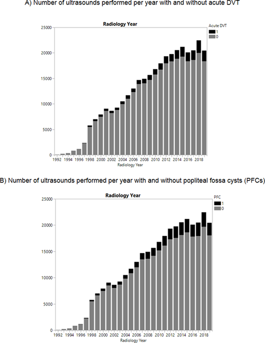
Results: A total of 332,016 lower extremity venous ultrasounds were performed in 223,035 patients; 156,846 unilateral and 175,170 bilateral lower extremities exams. The mean age at ultrasound was 63.3 (SD 16.5) and 54.7% were female. Ultrasound reports were available for analysis starting in 1992 with a significant increase in the number of ultrasounds performed over the study period across the enterprise (Figure 1). Overall, acute DVT was identified in 24,179 (7.3%) of ultrasounds, and PFCs were identified in 32,427 (9.8%) of ultrasounds. The sensitivity and specificity of the NLP algorithm in the full dataset to identify acute DVT was 86.0% and 97.2%, respectfully. The sensitivity and specificity of the NLP algorithm to identify PFCs was 97.8% and 99.5%, respectively. PFCs were present in 9.3% of ultrasounds with acute DVT and 9.8% of ultrasounds without acute DVT (p=0.007), OR 0.94 (95% CI 0.90-0.98). In a multivariate logistic regression model, after adjusting for age and sex, results remained significant (aOR 0.95, 95% CI 0.91-0.995). Comparing ultrasounds before and after 2010, there was a higher percentage of PFCs and acute DVT reported after 2010 (p<0.001 for both). Sensitivity analyses comparing results before or after 2010, by sex, and only in the first ultrasound performed per person, demonstrated similar results.
Conclusions: PFCs are negatively associated with the presence of acute DVT on lower extremity venous Duplex ultrasound. This data does not support PFCs as a contributing or causative factor in the development of lower extremity DVT.
No relevant conflicts of interest to declare.
Author notes
Asterisk with author names denotes non-ASH members.


This feature is available to Subscribers Only
Sign In or Create an Account Close Modal